Reading: Cell Structures and Function
http://www.ck12.org/saythanks
• Outline the structure of the plasma membrane.
• Distinguish cytoplasm from cytosol.
• Name three types of protein fibers that make up the cytoskeleton.
• Distinguish between cilia and flagella.
• Identify three structures that plant cells have but animal cells do not.
• List three major organelles found only in eukaryotic cells and identify their roles.
• Distinguish between a colonial organism and a multicellular organism.
• Outline the relationship between cells, tissues, organs, and organ systems.
Introduction
The invention of the microscope opened up a previously unknown world. Before the invention of the microscope, very little was known about what made up living things and non-living things, or where living things came from. During Hooke’s and Leeuwenhoek’s time, spontaneous generation —the belief that living organisms grow directly from decaying organic substances —was the accepted explanation for the appearance of small organisms. For example, people accepted that mice spontaneously appeared in stored grain, and maggots formed in meat with no apparent external influence. Once cells were discovered, the search for answers to such questions as "what are cells made of?" and "what do they do?" became the focus of study.
Cell Function
Cells share the same needs: the need to get energy from their environment, the need to respond to their environment, and the need to reproduce. Cells must also be able to separate their relatively stable interior from the ever-changing external environment. They do this by coordinating many processes that are carried out in different parts of the cell. Structures that are common to many different cells indicate the common history shared by cell-based life. Examples of these common structures include the components of both the cell (or plasma) membrane and the cytoskeleton, and other structures shown in Figure 1.1.
Plasma Membrane
The plasma membrane (also called the cell membrane) has many functions. For example, it separates the internal environment of the cell from the outside environment. It allows only certain molecules into and out of the cell. The ability to allow only certain molecules in or out of the cell is referred to as selective permeability or semipermeability. These semipermeable membranes regulate the cell’s interactions between the internal cytoplasm and the external surroundings. Proteins that are associated with the plasma membrane determine which molecules can pass through the membrane. This will be discussed in the next lesson. The plasma membrane also acts as the attachment point for both the intracellular cytoskeleton and, if present, the cell wall.
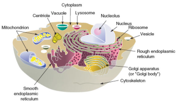
FIGURE 1.1
The structure and contents of a typical animal cell. Every animal cell has a cell membrane, cytoplasm, and a nucleus, but not all cells have every structure shown here. For example, some cells such as red blood cells do not have any mitochondria, yet others such as muscle cells may have thousands of mitochondria.
The plasma membrane is a lipid bilayer that is common to all living cells. A lipid bilayer is a double layer of closely-packed lipid molecules. The membranes of cell organelles are also lipid bilayers. The plasma membrane contains many different biological molecules, mostly lipids and proteins. These lipids and proteins are involved in many cellular processes.
Phospholipids
The main type of lipid found in the plasma membrane is phospholipid. A phospholipid is made up of a polar, phosphorus-containing head, and two long fatty acid, non-polar "tails." That is, the head of the molecule is hydrophilic (water-loving), and the tail is hydrophobic (water-fearing). Cytosol and extracellular fluid are made up of mostly water. In this watery environment, the water loving heads point out towards the water, and the water fearing tails point inwards, and push the water out. The resulting double layer is called a phospholipid bilayer. A phospholipid bilayer is made up of two layers of phospholipids, in which hydrophobic fatty acids are in the middle of the plasma membrane, and the hydrophilic heads are on the outside. An example of a simple phospholipid bilayer is illustrated in Figure 1.2.
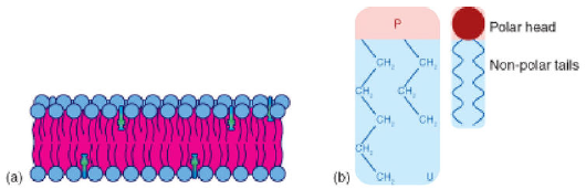
FIGURE 1.2
a) The hydrophobic fatty acids point towards the middle of the plasma membrane (pink), and the hydrophilic heads (blue) point outwards. The membrane is stabilized by cholesterol molecules (green). b) This self-organization of phospholipids results in a semipermeable membrane which allows only certain molecules in or out of the cell.
Plasma membranes of eukaryotes contain many proteins, as well as other lipids called sterols. The proteins have various functions, such as channels that allow certain molecules into the cell and receptors that bind to signal molecules. In Figure 1.2, the smaller (green) molecules shown between the phospholipids are cholesterol molecules. Cholesterol helps keep the plasma membrane firm and stable over a wide range of temperatures. At least ten different types of lipids are commonly found in plasma membranes. Each type of cell or organelle will have a different percentage of each lipid, protein and carbohydrate.
Membrane Proteins
Plasma membranes also contain certain types of proteins. A membrane protein is a protein molecule that is attached to, or associated with the membrane of a cell or an organelle. Membrane proteins can be put into two groups based on how the protein is associated with the membrane.
Integral membrane proteins are permanently embedded within the plasma membrane. They have a range of important functions. Such functions include channeling or transporting molecules across the membrane. Other integral proteins act as cell receptors. Integral membrane proteins can be classified according to their relationship with the bilayer:
• Transmembrane proteins span the entire plasma membrane. Transmembrane proteins are found
in all types of biological membranes.
• Integral monotopic proteins are permanently attached to the membrane from only one side.
Some integral membrane proteins are responsible for cell adhesion (sticking of a cell to another cell or surface). On the outside of cell membranes and attached to some of the proteins are carbohydrate chains that act as labels that identify the cell type. Shown in Figure 1.3 are two different types of membrane proteins and associated molecules.
Peripheral membrane proteins are proteins that are only temporarily associated with the membrane. They can be easily removed, which allows them to be involved in cell signaling. Peripheral proteins can also be attached to integral membrane proteins, or they can stick into a small portion of the lipid bilayer by themselves. Peripheral membrane proteins are often associated with ion channels and transmembrane receptors. Most peripheral membrane proteins are hydrophilic.
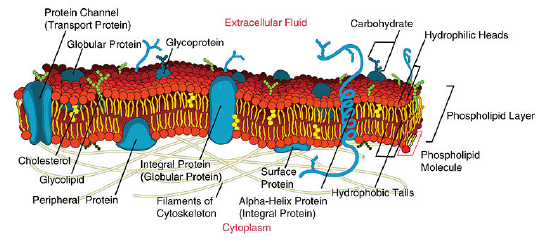
FIGURE 1.3
Some of the membrane proteins make up a major transport system that moves molecules and ions through the polar phospholipid bilayer.
Fluid Mosaic Model
In 1972 S.J. Singer and G.L. Nicolson proposed the now widely accepted Fluid Mosaic Model of the structure of cell membranes. The model proposes that integral membrane proteins are embedded in the phospholipid bilayer, as seen in Figure 1.3. Some of these proteins extend all the way through the bilayer, and some only partially across it. These membrane proteins act as transport proteins and receptors proteins. Their model also proposed that the membrane behaves like a fluid, rather than a solid. The proteins and lipids of the membrane move around the membrane, much like buoys in water. Such movement causes a constant change in the "mosaic pattern" of the plasma membrane.
Cytoplasm
The gel-like material within the cell that holds the organelles is called cytoplasm. The cytoplasm plays an important role in a cell, serving as a "jelly" in which organelles are suspended and held together by a fatty membrane. The cytosol, which is the watery substance that does not contain organelles, is made up of 80% to 90% water. The cytosol plays a mechanical role by exerting pressure against the cell’s plasma membrane which helps keep the shape of the cell. Cytosol also acts as the site of biochemical reactions such as anaerobic glycolysis and protein synthesis. In prokaryotes all chemical reactions take place in the cytosol.
Cytoskeleton
The cytoskeleton is a cellular "scaffolding" or "skeleton" that crisscrosses the cytoplasm. All eukaryotic cells have a cytoskeleton, and recent research has shown that prokaryotic cells also have a cytoskeleton. The eukaryotic cytoskeleton is made up of a network of long, thin protein fibers and has many functions. It helps to maintain cell shape. It holds organelles in place, and for some cells, it enables cell movement. The cytoskeleton also plays important roles in both the intracellular movement of substances and in cell division. Certain proteins act like a path that vesicles and organelles move along within the cell. The threadlike proteins that make up the cytoskeleton continually rebuild to adapt to the cell’s constantly changing needs. Three main kinds of cytoskeleton fibers are microtubules, intermediate filaments, and microfilaments.
• Microtubules, shown in Figure (a), are hollow cylinders and are the thickest of the cytoskeleton structures. They are most commonly made of filaments which are polymers of alpha and beta tubulin, and radiate outwards from an area near the nucleus called the centrosome. Tubulin is the protein that forms microtubules. Two forms of tubulin, alpha and beta, form dimers (pairs) which come together to form the hollow cylinders. The cylinders are twisted around each other to form the microtubules. Microtubules help the cell keep its shape. They hold organelles in place and allow them to move around the cell, and they form the mitotic spindle during cell division. Microtubules also make up parts of cilia and flagella, the organelles that help a cell move.
• Microfilaments, shown in Figure (b), are made of two thin actin chains that are twisted around one another. Microfilaments are mostly concentrated just beneath the cell membrane, where they support the cell and help the cell keep its shape. Microfilaments form cytoplasmatic extentions, such as pseudopodia and microvilli, which allow certain cells to move. The actin of the microfilaments interacts with the protein myosin to cause contraction in muscle cells. Microfilaments are found in almost every cell, and are numerous in muscle cells and in cells that move by changing shape, such as phagocytes (white blood cells that search the body for bacteria and other invaders).
• Intermediate filaments, shown in Figure (c), differ in make-up from one cell type to another. Intermediate filaments organize the inside structure of the cell by holding organelles and providing strength. They are also structural components of the nuclear envelope. Intermediate filaments made of the protein keratin are found in skin, hair, and nails cells.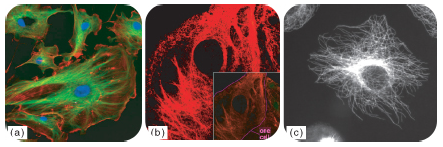
(a)The eukaryotic cytoskeleton. Microfilaments are shown in red, microtubules in green, and the nuclei are in blue. By linking regions of the cell together, the cytoskeleton helps support the shape of the cell. (b) Microscopy of keratin filaments (intermediate filaments) inside cells. (c) Microtubules in a methanol fixated cell, visualized with anti-beta-tubuline antibodies.
| Microtubules | Intermediate Filaments | Microfilaments | |
|---|---|---|---|
| Fiber Diameter | About 25 nm | 8 to 11 nm | Around 7 nm |
| Protein Composition | Tubulin, with two subunits, alpha and beta tubulin |
One of different types of proteins such as lamin, vimentin, and keratin |
Actin |
| Shape | Hollow cylinders made of two protein chains twisted around each other |
Protein fiber coils twisted into each other |
Two actin chains twisted around one another |
| Main Functions | Organelle and vesicle movement; form mitotic spindles during cell reproduction; cell motility (in cilia and flagella) |
Organize cell shape; positions organelles in cytoplasm structural
support of the nuclear envelope and sarcomeres; involved in cell-to-cell
and cell-tomatrix junctions |
Keep cellular shape; allows movement of certain cells by forming
cytoplasmatic extensions or contraction of actin fibers; involved in
some cell-to-cell or cell-to-matrix junctions |
| Image | Molecular structure of microtubules. |
Keratin intermediate filaments in skin cells (stained red). |
Actin cytoskeleton of mouse embryo cells. |
External Structures
Flagella (flagellum, singular) are long, thin structures that stick out from the cell membrane. Both eukaryotic and prokaryotic cells can have flagella. Flagella help single-celled organisms move or swim towards food. The flagella of eukaryotic cells are normally used for movement too, such as in the movement of sperm cells. The flagella of either group are very different from each other. Prokaryotic flagella, shown below, are spiral-shaped and stiff. They spin around in a fixed base much like a screw does, which moves the cell in a tumbling fashion. Eukaryotic flagella are made of microtubules and bend and flex like a whip.
Bacterial flagella spin about in place, which causes the bacterial cell to "tumble."
Cilia (cilium, singular) are made up of extensions of the cell membrane that contain microtubules. Although both are used for movement, cilia are much shorter than flagella. Cilia cover the surface of some single-celled organisms, such as paramecium. Their cilia beat together to move the little animals through the water. In multicellular animals, including humans, cilia are usually found in large numbers on a single surface of cells. Multicellular animals’ cilia usually move materials inside the body. For example, the mucociliary escalator of the respiratory system is made up of mucus-secreting cells that line the trachea and bronchi. Ciliated cells, shown in Figure 1.4, move mucus away
from the lungs. Spores, bacteria, and debris are caught in the mucus which is moved to the esophagus by the ciliated cells, where it is swallowed.
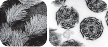
FIGURE 1.4
Left: Scanning electron micrograph (SEM), of the cilia sticking up from human lung cells. Right: Electron micrograph of cross-section of two cilia (not human), showing the positions of the microtubules inside. Note how there are nine groups of two microtubules (called dimers) in each cilium. Each dimer is made up of an alpha and a beta tubulin protein that are connected together.
The Nucleus and Other Organelles
The nucleus is a membrane-enclosed organelle found in most eukaryotic cells. The nucleus is the largest organelle in the cell and contains most of the cell’s genetic information (mitochondria also contain DNA, called mitochondrial DNA, but it makes up just a small percentage of the cell’s overall DNA content). The genetic information, which contains the information for the structure and function of the organism, is found encoded in DNA in the form of genes. A gene is a short segment of DNA that contains information to encode an RNA molecule or a protein strand. DNA in the nucleus is organized in long linear strands that are attached to different proteins. These proteins help the DNA to coil up for better storage in the nucleus. Think how a string gets tightly coiled up if you twist one end while holding the other end. These long strands of coiled-up DNA and proteins are called chromosomes. Each chromosome contains many genes. The function of the nucleus is to maintain the integrity of these genes and to control the activities of the cell by regulating gene expression. Gene expression is the process by which the information in a gene is "decoded" by various cell molecules to produce a functional gene product, such as a protein molecule or an RNA molecule.
The degree of DNA coiling determines whether the chromosome strands are short and thick or long and thin. Between cell divisions, the DNA in chromosomes is more loosely coiled and forms long thin strands called chromatin. Before the cell divides, the chromatin coil up more tightly and form chromosomes. Only chromosomes stain clearly enough to be seen under a microscope. The word chromosome comes from the Greek word chroma (color), and soma (body) due to its ability to be stained strongly by dyes.
Nuclear Envelope
The nuclear envelope is a double membrane of the nucleus that encloses the genetic material. It separates the contents of the nucleus from the cytoplasm. The nuclear envelope is made of two lipid bilayers, an inner membrane and an outer membrane. The outer membrane is continuous with the rough endoplasmic reticulum. Many tiny holes called nuclear pores are found in the nuclear envelope. These nuclear pores help to regulate the exchange of materials (such as RNA and proteins) between the nucleus and the cytoplasm.
Nucleolus
The nucleus of many cells also contains an organelle called a nucleolus, shown in Figure 1.5. The nucleolus is mainly involved in the assembly of ribosomes. Ribosomes are organelles made of protein and ribosomal RNA (rRNA), and they build cellular proteins in the cytoplasm. The function of the rRNA is to provide a way of decoding the genetic messages within another type of RNA called mRNA, into amino acids. After being made in the nucleolus, ribosomes are exported to the cytoplasm where they direct protein synthesis.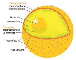
FIGURE 1.5
The eukaryotic cell nucleus. Visible in this diagram are the ribosome-studded double membranes of the nuclear envelope, the DNA (as chromatin), and the nucleolus. Within the cell nucleus is a viscous liquid called nucleoplasm, similar to the cytoplasm found outside the nucleus. The chromatin (which is normally invisible), is visible in this figure only to show that it is spread out throughout the nucleus.
Centrioles
Centrioles are rod-like structures made of short microtubules. Nine groups of three microtubules make up each centriole. Two perpendicularly placed centrioles make up the centrosome. Centrioles are very important in cellular division, where they arrange the mitotic spindles that pull the chromosome apart during mitosis.
Mitochondria
A mitochondrion (mitochondria, plural), is a membrane-enclosed organelle that is found in most eukaryotic cells. Mitochondria are called the "power plants" of the cell because they use energy from organic compounds to make ATP. ATP is the cell’s energy source that is used for such things such as movement and cell division. Some ATP is made in the cytosol of the cell, but most of it is made inside mitochondria. The number of mitochondria in a cell depends on the cell’s energy needs. For example, active human muscle cells may have thousands of mitochondria, while less active red blood cells do not have any.
As Figure 1.6 (a) and (b) shows, a mitochondrion has two phospholipids membranes. The smooth outer membrane separates the mitochondrion from the cytosol. The inner membrane has many folds, called cristae. The fluid-filled inside of the mitochondrian, called matrix, is where most of the cell’s ATP is made.
Although most of a cell’s DNA is contained in the cell nucleus, mitochondria have their own DNA. Mitochondria are able to reproduce asexually and scientists think that they are descended from prokaryotes. According to the endosymbiotic theory, mitochondria were once free-living prokaryotes that infected ancient eukaryotic cells. The invading prokaryotes were protected inside the eukaryotic host cell, and in turn the prokaryote supplied extra ATP to its host.
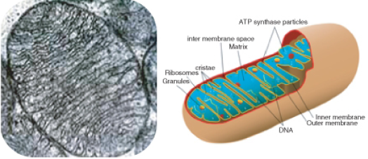
FIGURE 1.6
(a): Electron micrograph of a single mitochondrion within which you can see many cristae. Mitochondria range from 1 to 10 μm in size. (b): This model of a mitochondrian shows the organized arrangement of the inner and outer membranes, the protein matrix, and the folded inner mitochondrial membranes.
Endoplasmic Reticulum
The endoplasmic reticulum (ER) (plural, reticuli) is a network of phospholipid membranes that form hollow tubes, flattened sheets, and round sacs. These flattened, hollow folds and sacs are called cisternae. The ER has two major functions:
• Transport: Molecules, such as proteins, can move from place to place inside the ER, much like on an intracellular highway.
• Synthesis: Ribosomes that are attached to ER, similar to unattached ribosomes, make proteins. Lipids are also produced in the ER.
There are two types of endoplasmic reticulum, rough endoplasmic reticulum (RER) and smooth endoplasmic reticulum (SER).
• Rough endoplasmic reticulum is studded with ribosomes which gives it a "rough" appearance. These ribosomes make proteins that are then transported from the ER in small sacs called transport vesicles. The transport vesicles pinch off the ends of the ER. The rough endoplasmic reticulum works with the Golgi apparatus to move new proteins to their proper destinations in the cell. The membrane of the RER is continuous with the outer layer of the nuclear envelope.
• Smooth endoplasmic reticulum does not have any ribosomes attached to it, and so it has a smooth appearance. SER has many different functions some of which are: lipid synthesis, calcium ion storage, and drug detoxification. Smooth endoplasmic reticulum is found in both animal and plant cells and it serves different functions in each. The SER is made up of tubules and vesicles that branch out to form a network. In some cells there are dilated areas like the sacs of RER. Smooth endoplasmic reticulum and RER form an interconnected network.
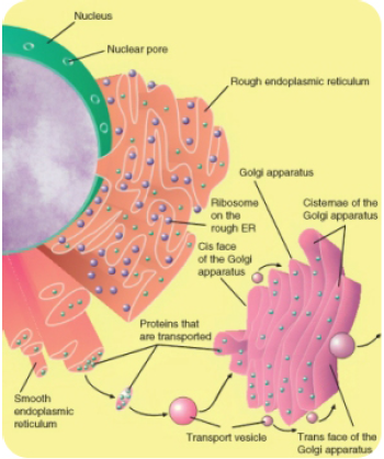
FIGURE 1.7
Image of nucleus, endoplasmic reticulum and Golgi apparatus, and how they work together. The process of secretion from endoplasmic reticuli (orange) to Golgi apparatus (pink) is shown.
Ribosomes
Ribosomes are small organelles and are the site of protein synthesis (or assembly). They are made of ribosomal protein and ribosomal RNA. Each ribosome has two parts, a large and a small subunit, as shown in Figure 1.8. The subunits are attached to each other. Ribosomes can be found alone or in groups within the cytoplasm. Some ribosomes are attached to the endoplasmic reticulum (as shown in Figure 1.7), and others are attached to the nuclear envelope.
Ribozymes are RNA molecules that catalyzes chemical reactions, such as translation. Translation is the process of ordering the amino acids in the assembly of a protein, and more will be discussed on translation in a later chapter. Briefly, the ribosomes interact with other RNA molecules to make chains of amino acids called polypeptide chains, due to the peptide bond that forms between individual amino acids. Polypeptide chains are built from the genetic instructions held within a messenger RNA molecule. Polypeptide chains that are made on the rough ER are inserted directly into the ER and then are transported to their various cellular destinations. Ribosomes on the rough ER usually produce proteins that are destined for the cell membrane.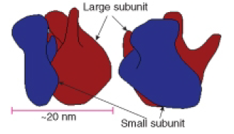
FIGURE 1.8
The two subunits that make up a ribosome, small organelles that are intercellular protein factories.
Golgi Apparatus
The Golgi apparatus is a large organelle that is usually made up of five to eight cup-shaped, membrane-covered discs called cisternae, as shown in Figure 1.7. The cisternae look a bit like a stack of deflated balloons. The Golgi apparatus modifies, sorts, and packages different substances for secretion out of the cell, or for use within the cell. The Golgi apparatus is found close to the nucleus of the cell where it modifies proteins that have been delivered in transport vesicles from the RER. It is also involved in the transport of lipids around the cell. Pieces of the Golgi membrane pinch off to form vesicles that transport molecules around the cell. The Golgi apparatus can be thought of as similar to a post office; it packages and labels "items" and then sends them to different parts of the cell. Both plant and animal cells have a Golgi apparatus. Plant cells can have up to several hundred Golgi stacks scattered throughout the cytoplasm. In plants, the Golgi apparatus contains enzymes that synthesize some of the cell wall polysaccharides.
Vesicles
A vesicle is a small, spherical compartment that is separated from the cytosol by at least one lipid bilayer. Many vesicles are made in the Golgi apparatus and the endoplasmic reticulum, or are made from parts of the cell membrane. Vesicles from the Golgi apparatus can be seen in Figure 1.7. Because it is separated from the cytosol, the space inside the vesicle can be made to be chemically different from the cytosol. Vesicles are basic tools of the cell for organizing metabolism, transport, and storage of molecules. Vesicles are also used as chemical reaction chambers. They can be classified by their contents and function.
• Transport vesicles are able to move molecules between locations inside the cell. For example,
transport vesicles move proteins from the rough endoplasmic reticulum to the Golgi apparatus.
• Lysosomes are vesicles that are formed by the Golgi apparatus. They contain powerful enzymes
that could break down (digest) the cell. Lysosomes break down harmful cell products, waste
materials, and cellular debris and then force them out of the cell. They also digest invading
organisms such as bacteria. Lysosomes also break down cells that are ready to die, a process
called autolysis.
• Peroxisomes are vesicles that use oxygen to break down toxic substances in the cell. Unlike
lysosomes, which are formed by the Golgi apparatus, peroxisomes self replicate by growing
bigger and then dividing. They are common in liver and kidney cells that break down harmful
substances. Peroxisomes are named for the hydrogen peroxide (H2O2) that is produced when
they break down organic compounds. Hydrogen peroxide is toxic, and in turn is broken down
into water (H2O) and oxygen (O2) molecules.
Vacuoles
Vacuoles are membrane-bound organelles that can have secretory, excretory, and storage functions. Many organisms will use vacuoles as storage areas and some plant cells have very large vacuoles. Vesicles are much smaller than vacuoles and function in transporting materials both within and to the outside of the cell.
Special Structures in Plant Cells
Most of the organelles that have been discussed are common to both animal and plant cells. However, plant cells also have features that animal cells do not have; they have a cell wall, a large central vacuole, and plastids such as chloroplasts.
Plants have very different lifestyles from animals, and these differences are apparent when you examine the structure of the plant cell. Plants make their own food in a process called photosynthesis. They take in carbon dioxide (CO2) and water (H2O) and convert them into sugars. The features unique to plant cells can be seen in Figure 1.9.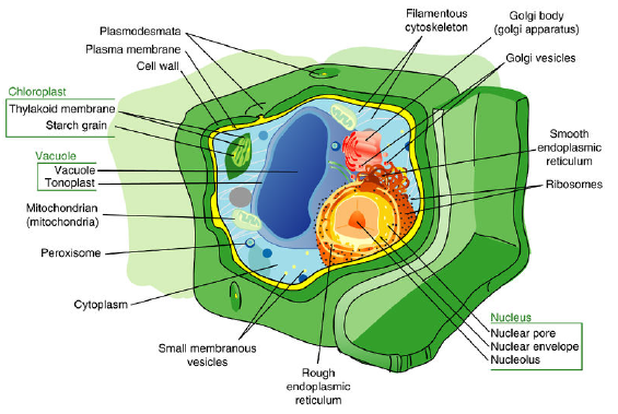
FIGURE 1.9
In addition to containing most of the organelles found in animal cells, plant cells also have a cell wall, a large central vacuole, and plastids. These three features are not found in animal cells.
Cell Wall
A cell wall is a rigid layer that is found outside the cell membrane and surrounds the cell. The cell wall contains not only cellulose and protein, but other polysaccharides as well. In fact, two other classes of polysaccharides, hemicelluloses and pectic polysaccharides, can comprise 30% of the dry mass of the cell wall. The cell wall provides structural support and protection. Pores in the cell wall allow water and nutrients to move into and out of the cell. The cell wall also prevents the plant cell from bursting when water enters the cell.
Microtubules guide the formation of the plant cell wall. Cellulose is laid down by enzymes to form the primary cell wall. Some plants also have a secondary cell wall. The secondary wall contains a lignin, a secondary cell component in plant cells that have completed cell growth/expansion.
Central Vacuole
Most mature plant cells have a central vacuole that occupies more than 30% of the cell’s volume, but can also occupy as much as 90% of the volume of certain cells. The central vacuole is surrounded by a membrane called the tonoplast. The central vacuole has many functions. Aside from storage, the main role of the vacuole is to maintain turgor pressure against the cell wall. Proteins found in the tonoplast control the flow of water into and out of the vacuole. The central vacuole also stores the pigments that color flowers.
The central vacuole contains large amounts of a liquid called cell sap, which differs in composition to the cell cytosol. Cell sap is a mixture of water, enzymes, ions, salts, and other substances. Cell sap may also contain toxic byproducts that have been removed from the cytosol. Toxins in the vacuole may help to protect some plants from being eaten.
Plastids
Plant plastids are a group of closely related membrane-bound organelles that carry out many functions. They are responsible for photosynthesis, for storage of products such as starch, and for the synthesis of many types of molecules that are needed as cellular building blocks. Plastids have the ability to change their function between these and other forms. Plastids contain their own DNA and some ribosomes, and scientists think that plastids are descended from photosynthetic bacteria that allowed the first eukaryotes to make oxygen. The main types of plastids and their functions are:
• Chloroplasts are the organelle of photosynthesis. They capture light energy from the sun and use it with water and carbon dioxide to make food (sugar) for the plant. The arrangement of chloroplasts in a plant’s cells can be seen in Figure 1.10.
• Chromoplasts make and store pigments that give petals and fruit their orange and yellow colors.
• Leucoplasts do not contain pigments and are located in roots and non-photosynthetic tissues of plants. They may become specialized for bulk storage of starch, lipid, or protein. However, in many cells, leucoplasts do not have a major storage function; instead they make molecules such as fatty acids and many amino acids.
Chloroplasts capture light energy from the sun and use it with water and carbon dioxide to produce sugars for food. Chloroplasts look like flat discs that are usually 2 to 10 micrometers in diameter and 1 micrometer thick. A model of a chloroplast is shown in Figure 1.11. The chloroplast is enclosed by an inner and an outer phospholipid membrane. Between these two layers is the intermembrane space. The fluid within the chloroplast is called the stroma, and it contains one or more molecules of small circular DNA. The stroma also has ribosomes. Within the stroma are stacks of thylakoids, the sub organelles which are the site of photosynthesis. The thylakoids are arranged in stacks called
grana (singular: granum). A thylakoid has a flattened disk shape. Inside it is an empty area called the thylakoid space or lumen. Photosynthesis takes place on the thylakoid membrane.
Within the thylakoid membrane is the complex of proteins and light-absorbing pigments, such as chlorophyll and carotenoids. This complex allows capture of light energy from many wavelengths because chlorophyll and carotenoids both absorb different wavelengths of light. You will learn more about how chloroplasts convert light energy into chemical energy in the Photosynthesis chapter.
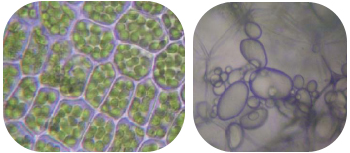
FIGURE 1.10
Plant cells with visible chloroplasts (left). Starch-storing potato leucoplasts (right).
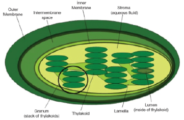
FIGURE 1.11
The internal structure of a chloroplast, with a granal stack of thylakoids circled.
Organization of Cells
Biological organization exists at all levels in organisms. It can be seen at the smallest level, in the molecules that made up such things as DNA and proteins, to the largest level, in an organism such as a blue whale, the largest mammal on Earth. Similarly, single celled prokaryotes and eukaryotes show order in the way their cells are arranged. Single-celled organisms such as an amoeba are free-floating and independent-living. Their single-celled "bodies" are able to carry out all the processes of life such as metabolism and respiration without help from other cells. Some single-celled organisms such as bacteria can group together and form a biofilm. A biofilm is a large grouping of many bacteria that sticks to a surface and makes a protective coating over itself. Biofilms can show similarities to multicellular organisms. Division of labor is the process in which one group of cells does one job (such as making the "glue" that sticks the biofilm to the surface) while another group of cells does another job (such as taking in nutrients). Multicellular organisms carry out their life processes through division of labor and they have specialized cells that do specific jobs. However, biofilms are not considered a multicellular organism and are instead called colonial organisms. The difference between a multicellular organism and a colonial organism is that individual organisms from a colony or biofilm can, if separated, survive on their own, while cells from a multicellular organism (e.g., liver cells) cannot.
Colonial Organisms
Colonial organisms were probably one of the first evolutionary steps towards multicellular organisms. Algae of the genus Volvox are an example of the border between colonial organisms and multicellular organisms.
Each Volvox is a colonial organism. It is made up of between 1000 to 3000 photosynthetic algae that are grouped together into a hollow sphere. The sphere has a distinct front and back end. The cells have eyespots, which are more developed in the cells near the front. This enables the colony to swim towards light.
Origin of Multicellularity
The oldest known multicellular organism is a red algae Bangiomorpha pubescens, fossils of which were found in 1.2 billion year old rock. However, the first organisms were single celled. How multicellular organisms developed is the subject of much debate.
Scientists think that multicellularity arose from cooperation between many organisms of the same species. The Colonial Theory proposes that this cooperation led to the development of a multicellular organism. Many examples of cooperation between organisms in nature have been observed. For example, a certain species of amoeba (a single-celled animal) groups together during times of food shortage and forms a colony that moves as one to a new location. Some of these amoebas then become slightly differentiated from each other. Volvox is another example of a colonial organism. Most scientists accept that the Colonial theory explains how multicellular organisms evolved.
Multicellular organisms are organisms that are made up of more than one type of cell and have specialized cells that are grouped together to carry out specialized functions. Most life that you can see without a microscope is multicellular. As discussed earlier, the cells of a multicellular organism would not survive as independent cells. The body of a multicellular organism, such as a tree or a cat, exhibits organization at several levels: tissues, organs, and organ systems. Similar cells are grouped into tissues, groups of tissues make up organs, and organs with a similar function are grouped into an organ system.
Levels of Organization in Multicellular Organisms
The simplest living multicellular organisms, sponges, are made of many specialized types of cells that work together for a common goal. Such cell types include digestive cells, tubular pore cells; and epidermal cells. Though the different cell types create a large organized, multicellular structure—the visible sponge—they are not organized into true interconnected tissues. If a sponge is broken up by passing it through a sieve, the sponge will reform on the other side. However, if the sponge’s cells are separated from each other, the individual cell types cannot survive alone. Simpler colonial organisms, such as members of the genus Volvox differ in that their individual cells are free-living and can survive on their own if separated from the colony.
A tissue is a group of connected cells that have a similar function within an organism. More complex organisms such as jellyfish, coral, and sea anemones have a tissue level of organization. For example, jellyfish have tissues that have separate protective, digestive, and sensory functions.
Even more complex organisms, such as the roundworm, while also having differentiated cells and tissues, have an organ level of development. An organ is a group of tissues that has a specific function or group of functions. Organs can be as primitive as the brain of a flatworm (a group of nerve cells), as large as the stem of a sequoia (up to 90 meters, or 300 feet, in height), or as complex as a human liver.
The most complex organisms (such as mammals, trees, and flowers) have organ systems. An organ system is a group of organs that act together to carry out complex related functions, with each organ focusing on a part of the task. An example is the human digestive system in which the mouth ingests food, the stomach crushes and liquifies it, the pancreas and gall bladder make and release digestive enzymes, and the intestines absorb nutrients into the blood.
Lesson Summary
• The plasma membrane is a selectively permeable lipid bilayer that contains mostly lipids and proteins. These lipids and proteins are involved in many cellular processes.
• The gel-like material within the cell that holds the organelles is called cytoplasm. The cytosol, which is the watery substance that does not contain organelles, is made up of 80% to 90% water.
• The cytoskeleton has many functions. It helps to maintain cell shape, it holds organelles in place, and for some cells, it enables cell movement. The cytoskeleton also plays important roles in both the intracellular movement of substances and in cell division. Three main kinds of cytoskeleton fibers are microtubules, intermediate filaments, and microfilaments.
• Cilia are extensions of the cell membrane that contain microtubules. Although both are used for movement, cilia are much shorter than flagella. Cilia cover the surface of some single-celled animals, such as paramecium, but cover only one side of cells in some multicellular organisms.
• There are three features that plant cells have that animal cells do not have: a cell wall, a large central vacuole, and plastids.
• Mitochondria use energy from organic compounds to make ATP.
• Ribosomes are exported from the nucleolus, where they are made, to the cytoplasm.
• The Golgi apparatus is a large organelle that is usually made up of five to eight cup-shaped, membrane-covered discs called cisternae. It modifies, sorts, and packages different substances for secretion out of the cell, or for use within the cell.
• Individual organisms from a colonial organism or biofilm can, if separated, survive on their own, while cells from a multicellular organism (e.g., liver cells) cannot.
• A tissue is a group of connected cells that have a similar function within an organism. An organ is a group of tissues that has a specific function or group of functions, and an organ system is a group of organs that act together to perform complex related functions, with each organ focusing on a part of the task.
Review Questions
1. What are the main components of a plasma membrane?
2. What does the fluid mosaic model describe?
3. What is the difference between cytoplasm and cytosol?
4. What type of molecule is common to all three parts of the cytoskeleton?
5. Name the three main parts of the cytoskeleton.
6. What structures do plant cells have that animal cells do not have?
7. Identify two functions of plastids in plant cells.
8. What is the main difference between rough endoplasmic reticulum and smooth endoplasmic reticulum?
9. List five organelles eukaryotes have that prokaryotes do not have.
10. What is a cell feature that distinguishes a colonial organism from a multicellular organism?
11. What is the difference between a cell and a tissue?
12. Identify two functions of the nucleus.
13. Identify the reason why mitochondria are called "power plants" of the cell.
14. If muscle cells become more active than they usually are, they will grow more mitochondria. Explain why this happens.
Vocabulary
chloroplast: The organelle of photosynthesis; captures light energy from the sun and uses it with water and carbon dioxide to make food (sugar) for the plant.
cilia (cilium): Made up of extensions of the cell membrane that contain microtubules; involved in movement.
cell wall: A rigid layer that is found outside the cell membrane and surrounds the cell; provides structural support and protection.
cytoplasm: The gel-like material within the cell that holds the organelles.
cytoskeleton: A cellular "scaffolding" or "skeleton" that crisscrosses the cytoplasm; helps to maintain cell shape, it holds organelles in place, and for some cells, it enables cell movement.
endoplasmic reticulum (ER): A network of phospholipid membranes that form hollow tubes, flattened sheets, and round sacs; involved in transport of molecules, such as proteins, and the synthesis of proteins and lipids.
flagella (flagellum): Long, thin structures that stick out from the cell membrane; help single-celled organisms move or swim towards food.
Fluid Mosaic Model: Model of the structure of cell membranes; proposes that integral membrane proteins are embedded in the phospholipid bilayer; some of these proteins extend all the way through the bilayer, and some only partially across it; also proposes that the membrane behaves like a fluid, rather than a solid.
gene: A short segment of DNA that contains information to encode an RNA molecule or a protein strand.
gene expression: The process by which the information in a gene is "decoded" by various cell molecules to produce a functional gene product, such as a protein molecule or an RNA molecule.
Golgi apparatus: A large organelle that is usually made up of five to eight cup-shaped, membrane-covered discs called cisternae; modifies, sorts, and packages different substances for secretion out of the cell, or for use within the cell.
integral membrane proteins: Proteins that are permanently embedded within the plasma membrane; involved in channeling or transporting molecules across the membrane or acting as cell receptors.
intermediate filaments: Filaments that organize the inside structure of the cell by holding organelles and providing strength.
lipid bilayer: A double layer of closely-packed lipid molecules; the cell membrane is a phospholipid bilayer.
lysosome: A vesicle that contains powerful digestive enzymes.
membrane protein: A protein molecule that is attached to, or associated with the membrane of a cell or an organelle.
microfilament: Filament made of two thin actin chains that are twisted around one another; organizes cell shape; positions organelles in cytoplasm; involved in cell-to-cell and cell-to-matrix junctions.
microtubules: Hollow cylinders that make up the thickest of the cytoskeleton structures; made of the protein tubulin, with two subunits, alpha and beta tubulin; involved in organelle and vesicle movement; form mitotic spindles during cell division; involved in cell motility (in cilia and flagella).
mitochondria (mitochondrion): Membrane-enclosed organelles that are found in most eukaryotic cells; called the "power plants" of the cell because they use energy from organic compounds to make ATP.
multicellular organisms: Organisms that are made up of more than one type of cell; have specialized cells that are grouped together to carry out specialized functions.
nucleus: The membrane-enclosed organelle found in most eukaryotic cells; contains the genetic material (DNA).
organ: A group of tissues that has a specific function or group of functions.
organ system: A group of organs that acts together to carry out complex related functions, with each organ focusing on a part of the task.
peripheral membrane proteins: Proteins that are only temporarily associated with the membrane; can be easily removed, which allows them to be involved in cell signaling.
peroxisomes: Vesicles that use oxygen to break down toxic substances in the cell.
phospholipid: A lipid made up of up of a polar, phosphorus-containing head, and two long fatty acid, non-polar "tails." The head of the molecule is hydrophilic (water-loving), and the tail is hydrophobic (water-fearing).
plasma membrane: Phospholipid bilayer that separates the internal environment of the cell from the outside environment.
ribosomes: Organelles made of protein and ribosomal RNA (rRNA); where protein synthesis occurs.
selective permeability: The ability to allow only certain molecules in or out of the cell; characteristic of the cell membrane; also called the cell membrane.
spontaneous generation: The belief that living organisms grow directly from decaying organic substances.
tissue: A group of connected cells that has a similar function within an organism.
transport vesicle: A vesicle that is able to move molecules between locations inside the cell.
vacuole: Membrane-bound organelles that can have secretory, excretory, and storage functions; plant cells have a large central vacuole
vesicle: A small, spherical compartment that is separated from the cytosol by at least one lipid bilayer.
Points to Consider
• How do you think small molecules, or even water, get through the cell membrane?
• Is it possible that proteins help in this transport process?
• What type of proteins would help with transport?
Last modified: Monday, August 13, 2018, 8:28 AM